Medical Image Segmentation
In the rapidly evolving field of healthcare, Artificial Intelligence (AI) has paved the way for revolutionary advancements. One such breakthrough is medical image segmentation, where precise delineation of structures within medical images is crucial for diagnostics and treatment planning. This article discusses my project, which involved comparing two cutting-edge deep learning architectures—U-Net and Duck-Net—and their effectiveness in improving medical image segmentation.
The Role of Medical Image Segmentation
Medical image segmentation is a crucial aspect of computer-aided diagnosis, particularly in procedures like colonoscopy, where accurate detection of polyps can be life-saving. In this study, I conducted a detailed comparative analysis of two powerful deep learning architectures (U-Net and Duck-Net) focusing on their performance in polyp segmentation within colonoscopy images. Using the CVC-ClinicDB dataset, this project aims to enhance the precision of polyp detection, ultimately contributing to advancements in medical image analysis.
Dataset and Methodology
For this research, I utilized the CVC-ClinicDB dataset, which contains 612 meticulously annotated images specifically curated for polyp detection. Each image in the dataset comes with precise polyp location and size information, making it a robust foundation for training and evaluating segmentation models.
The core of the research lies in the application of two convolutional neural network (CNN) architectures: U-Net, a widely acclaimed model for medical image segmentation, and Duck-Net, a more recent innovation that features advanced convolutional blocks designed for finer feature extraction. Both models were trained using techniques such as data augmentation, adaptive learning rate scheduling, and comprehensive preprocessing of images.
Model Architecture and Development
U-Net was chosen due to its powerful encoder-decoder architecture, which is particularly effective for retaining both local and global features within medical images. Its skip connections between the encoder and decoder parts of the network allow it to maintain crucial image details during segmentation.
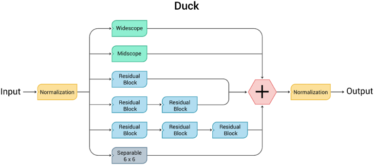
Duck-Net, on the other hand, introduces a unique “duckv2” convolutional block that significantly enhances feature extraction across multiple scales. This enables Duck-Net to outperform traditional models in detecting fine-grained details, making it a superior choice for complex segmentation tasks.
Results and Performance Metrics
The performance of both architectures was evaluated using metrics such as the Dice Coefficient, Intersection over Union (IoU), Precision, Recall, and Accuracy. The results of the study showed that Duck-Net consistently outperformed U-Net, achieving a higher Dice Coefficient (0.9208 compared to U-Net’s 0.8581) and IoU, particularly in challenging cases with irregular polyp shapes or sizes. However, U-Net demonstrated sporadic successes, showing that it remains a reliable tool for specific tasks.

Conclusion
This project highlights the strengths and weaknesses of both U-Net and Duck-Net in medical image segmentation, with Duck-Net showing a clear edge in polyp detection. While U-Net remains a solid performer in certain scenarios, Duck-Net’s advanced convolutional blocks make it a more suitable choice for complex segmentation tasks. These findings pave the way for future research in AI-driven medical imaging, offering a potential roadmap for more accurate and efficient diagnostic tools in healthcare.
Complete Report
A complete report of this research can be found and downloaded here or can be seen below.